Imaging in PRP Injections: Ultrasound and Fluoroscopy Compared

Precise PRP delivery: what ultrasound and fluoroscopy can do

Professional information for physicians and medical professionals. This article reviews the current evidence and does not replace guidelines or individual medical decision-making. Research status: September 2026.

Image-guided PRP injection

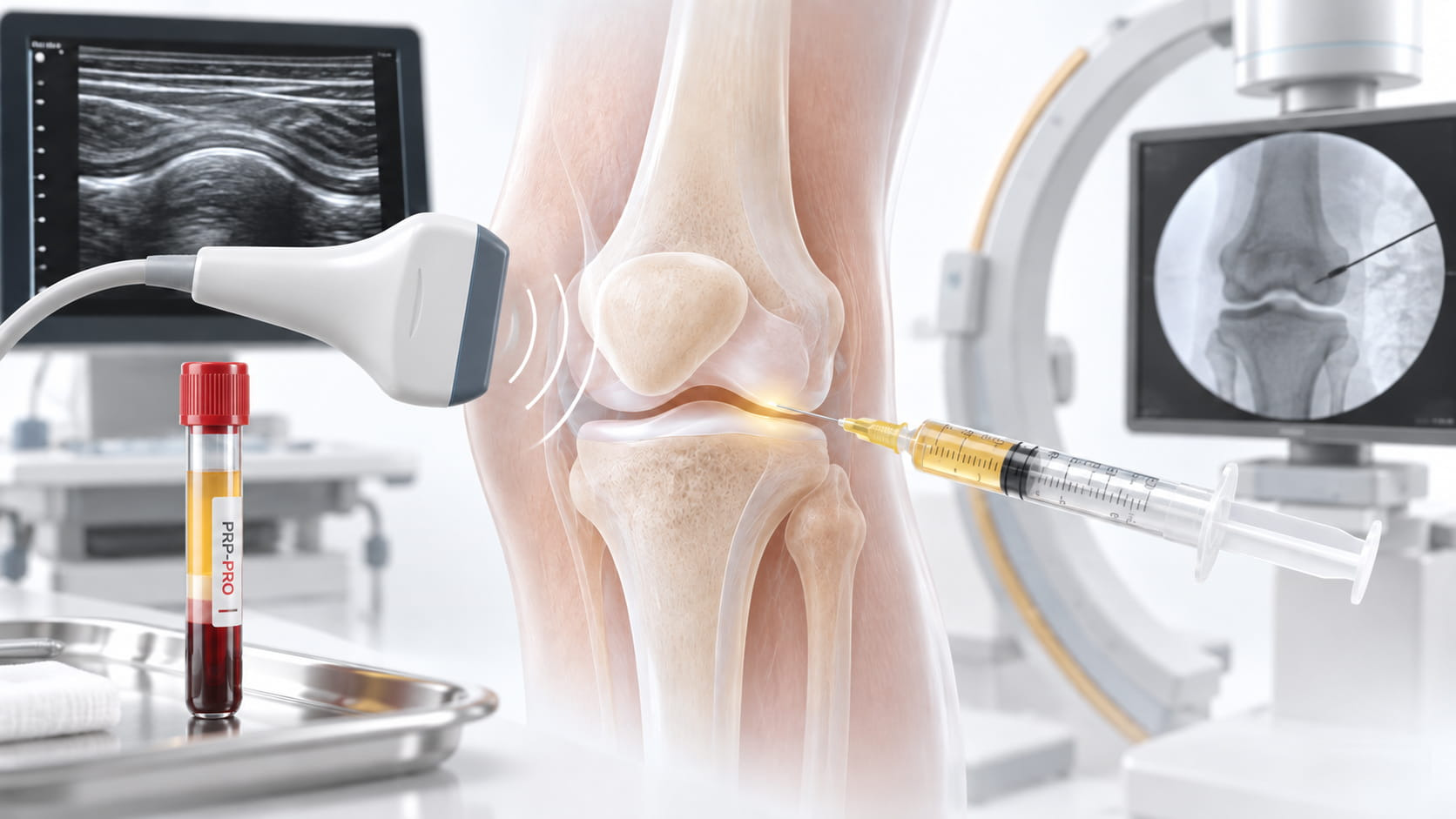
For small, deep or anatomically confined target spaces, the position of the needle tip can determine whether a preparation is actually delivered where intended. Ultrasound and fluoroscopy monitor this position in different ways.

With an ultrasound-guided PRP injection soft tissues, the needle and adjacent vessels or nerves can be visualised in real time. A fluoroscopy-guided PRP injection is oriented primarily to bony structures; contrast medium can additionally confirm the position in joint, disc or epidural spaces.

The key scientific distinction is this: image guidance can increase targeting accuracy . Whether this also improves the clinical outcome of a PRP treatment is a separate question and has so far been studied directly for only a few indications.

Out of 100 injections, how many reach the joint …

Select an approach or imaging method.

Knee, without image guidance

Sacroiliac joint, with image guidance

75 %intra-articular

Anteromedial approach without image guidance; position checked fluoroscopically. Jackson et al. 2002

Hits: landmark guidance Hits: image guidance outside the joint

Rounded representation of published accuracy rates1,2,8. The values come from different studies using corticosteroid, hyaluronic acid or contrast-medium injections and do not indicate the efficacy of PRP.

Summary

The key points in four statements

Accuracy depends on the target and approach.At the knee, favourable landmark-guided approaches achieve high accuracy rates, while others perform substantially worse. Image guidance improves accuracy in many musculoskeletal interventions.24

Not all imaging is the same.EULAR favours ultrasound for targeted interventions at peripheral joints and nerves. For the spine and sacroiliac joint, the modality should be selected according to the target structure, procedure, experience, availability and radiation exposure.25

Direct PRP comparisons exist – but only a few.In lateral epicondylitis, a randomised study found no clinical advantage of ultrasound guidance; a small randomised temporomandibular-joint study from 2026, by contrast, found greater placement accuracy and better trajectories for some outcomes.26,27

Precision is not the same as efficacy.A correctly placed injection does not prove that PRP is effective for the respective indication. Preparation, application and clinical effect must be assessed separately.

Background

Why the injection site matters particularly in PRP

Greater anatomical precision does not automatically lead to a better clinical result with every injectate. In a double-blind rotator-cuff study, for example, an ultrasound-guided subacromial steroid injection was not significantly more effective than the same dose given systemically by intramuscular injection7. Such steroid data cannot be transferred to PRP, but they show why placement accuracy and clinical outcome must be studied separately.

For PRP, a local effect is biologically plausible: platelets and released mediators are initially present where the preparation is applied. This does not automatically mean, however, that every millimetre-perfect placement is clinically superior. So far, this question has been examined in only a few direct PRP comparison studies.26,27

Imaging can also provide information beyond the needle position itself: ultrasound can show effusions, tendon structure, vessels and nerves, for example. An effusion changes volume and distribution within a joint; this alone does not prove that aspiration before PRP improves the clinical outcome. The quality of PRP preparation and the quality of application remain two separate process steps.

Accuracy

What landmark-guided injections actually hit

The most frequently cited knee study is by Jackson and colleagues. An experienced orthopaedic surgeon performed 240 injections into knees without effusion, 80 through each of three approaches, and subsequently checked the position fluoroscopically with contrast medium. The intra-articular rates were 93 percent for the lateral mid-patellar approach, 75 percent for the anteromedial approach and 71 percent for the anterolateral approach1.

A systematic review of 23 publications confirms the pattern2. Pooled data showed the superolateral approach without image guidance to be the most accurate at 87 percent, while the medial mid-patellar approach was least accurate at 64 percent. Overall, roughly one in five blind knee injections was extra-articular. Image-guided injections were moderately more accurate, particularly with less favourable approaches. Experienced injectors achieved good results without imaging using the superolateral approach.

The deeper and more obscured by bone the target, the larger the difference can become. At the sacroiliac joint, a prospective randomised study directly compared ultrasound and fluoroscopy: 87.3 percent of ultrasound-guided and 98.2 percent of fluoroscopy-guided injections were intra-articular8.

A broader systematic EULAR review of 66 studies, including 49 randomised trials, points in the same direction: image-guided procedures were more accurate than palpation or anatomical landmarks in many comparisons. The authors nevertheless emphasised study heterogeneity and frequently moderate-to-high risk of bias; direct comparisons of imaging methods did not identify one universally superior modality.24

The American Medical Society for Sports Medicine also summarised the literature in 2015. Ultrasound-guided injections were rated as more accurate than landmark-guided injections (SORT A), while evidence for efficacy and cost-effectiveness was weaker (SORT B).3 The newer EULAR Points to Consider therefore take a target-specific approach: ultrasound is preferred for peripheral joints and nerves, while the imaging modality for the spine and sacroiliac joint should be selected individually.25

Interpretation

Accuracy is not the same as efficacy

Whether the needle reaches the intended structure is a technical endpoint. Whether the patient gains additional clinical benefit is a different endpoint. This distinction is central to PRP.

A direct randomised PRP study in chronic lateral epicondylitis compared 30 palpation-guided with 30 ultrasound-guided injections. Pain, DASH score and grip strength improved in both groups; there was no significant difference between the techniques after one, three or six months.26

A small randomised temporomandibular-joint study from 2026 produced a different result. In 64 patients, placement after the PRP injection was checked fluoroscopically with contrast medium. Ultrasound achieved 32 of 32 correct placements, compared with 22 of 32 with palpation (100% vs. 68.8%). For some pain and functional parameters, the ultrasound group showed advantages after three or six months.27 Because of the small sample, the single operator and the specific target region, this does not establish a general superiority of ultrasound-guided PRP.

PRP studies in which imaging is standardised in both groups also underline the need to separate application from efficacy. In chronic Achilles tendinopathy, ultrasound-guided PRP was no better than the respective control treatment in randomised studies.10,11 Precise application can reduce one methodological source of error, but it does not replace evidence of efficacy for the indication.

Direct PRP data: available, but limited

The statement that “there are no direct comparison studies” is no longer tenable. Randomised data exist at least for lateral epicondylitis and the temporomandibular joint, with differing results.26,27 This is not enough for a robust, indication-independent conclusion. Standardised reporting of the PRP preparation and application also remains important; the MIBO checklist was developed in part for this purpose.20

Ultrasound

Ultrasound: real-time imaging for soft tissues and peripheral joints

High-frequency linear probes are used for superficial targets such as tendons, knee, elbow and shoulder. They provide fine resolution but lose image quality with increasing depth. For the hip and for patients with a greater soft-tissue depth, lower-frequency curved-array probes penetrate more deeply at the cost of lower resolution.

Two techniques are distinguished for needle guidance. In-Plane the needle runs within the ultrasound plane and can be seen along its full length. Out-of-Plane the needle crosses the plane and appears only as a bright point, making it easy to mistake the shaft for the tip. The steeper the insertion angle, the less ultrasound is reflected back from the needle to the probe.

The practical advantage is the moving image: the target structure and needle can be observed during advancement, and distribution of the injectate can often be followed sonographically depending on the tissue and target space. If the observed distribution pattern does not match the intended target, the position can be reassessed. Doppler techniques can also show vessels along the planned needle path.

For peripheral joints and nerves, the EULAR Points to Consider favour ultrasound over other imaging modalities.25 The widespread use of ultrasound in PRP research is also shown by a systematic review of tendinopathies including 33 randomised studies and 2,025 participants.28 These studies do not automatically prove that ultrasound itself causes the PRP effect.

Strengths

  • No ionising radiation; repeated real-time monitoring is possible
  • Real-time visualisation of injectate distribution
  • Effusion, tendon lesion, vessels and nerves in the same image
  • Suitable for office-based use, with comparatively modest investment

Limitations

  • Highly operator-dependent; training, knowledge of regional anatomy and practical experience are crucial
  • No view through or inside bone
  • Image quality decreases with depth and obesity
  • At the sacroiliac joint, placement in the dorsal ligamentous structures is more frequent than intra-articular placement8

Fluoroscopy

Fluoroscopy: contrast medium shows what PRP does not

Under a C-arm, the needle and bony landmarks are directly visible. In many intra-articular and spinal interventions, a small amount of iodinated contrast medium is additionally used to assess the distribution pattern in the intended target space and to identify signs of unintended vascular or intrathecal placement. Contrast medium therefore supplements anatomical needle control; it is not required in the same way for every fluoroscopic procedure.

One important point: PRP is not radiopaque and therefore cannot be seen directly under fluoroscopy as its own contrast agent. If contrast medium is used beforehand, its pattern confirms the position or accessible target space; the subsequent distribution of PRP itself is not directly visualised by this.

PRP-specific fluoroscopy data come mainly from axial and deep target regions. In discogenic low-back pain, a small double-blind randomised trial reported short-term advantages after intradiscal PRP compared with the control group.13 For lumbar intradiscal and epidural PRP procedures, the 2025 ASIPP guideline rates the evidence as Level III (“fair”), and for facet and sacroiliac joints as Level IV (“limited”).30

The sacroiliac joint also illustrates why technical precision and efficacy must remain separate: in a double-blind fluoroscopy-guided RCT with 26 patients, both PRP and corticosteroid groups improved, but PRP was not superior to steroid.29 A 2026 meta-analysis of ten randomised or quasi-randomised studies on facet and SI-joint pain found some later pain outcomes favouring PRP, but still judged the overall evidence inconclusive.31

CT can be an alternative for selected spinal interventions, such as interlaminar epidural PRP injections.15 Conversely, ultrasound has also been studied in a randomised trial for transforaminal PRP injections.21 The methods are therefore not universally interchangeable; target structure, intervention, experience, equipment availability and radiation exposure should be considered together.25

Radiation protection in Germany

For the medical use of ionising radiation, a justified indication by a physician or dentist with the required radiation-protection expertise is mandatory (§ 83 StrlSchG). The Radiation Protection Ordinance further specifies, among other things, the assessment of the procedure or a treatment attempt requiring special justification and the requirements for technical performance (§§ 119, 145 StrlSchV).22 Exposure must be limited as far as medically reasonable.

Target structure

Which imaging method fits which target structure?

The choice depends on more than depth. The target structure, planned procedure, anatomical overlap, operator experience, equipment availability and, for X-ray procedures, radiation exposure all interact.25 The following assignment is therefore a practical orientation, not a procedural requirement.

Soft tissues and peripheral joints

Axial skeleton and bone

Location of target structure: Superficial

Knee joint

Ultrasound, linear probeWithout image guidance: superolateral is most accurate

Why this method

The suprapatellar recess is readily visualised. An effusion can be identified and, when clinically indicated, aspirated before PRP administration; whether this improves outcomes is a separate question.

Evidence

Without image guidance, roughly one in five injections was extra-articular; image-guided injections were moderately more accurate, especially with medial mid-patellar and anterolateral approaches2.

Pitfall

In a dry osteoarthritic knee, an effusion is unavailable as a landmark. Anteromedial and anterolateral approaches are substantially less reliable without imaging than the lateral mid-patellar approach1.

Evidence

The evidence at a glance

Filter by imaging modality and study type. Note the label at the top right: many statements on accuracy do not come from PRP studies.

Imaging

Study type

Loading study overview

Jackson et al. 2002Accuracy

240 knee injections without effusion, one investigator, position checked fluoroscopically

Intra-articular: lateral mid-patellar 93%, anteromedial 75%, anterolateral 71%.1

KneeWithout image guidance
Maricar et al. 2013Accuracy

Systematic review, 23 publications

Without imaging, roughly one in five injections was extra-articular; image guidance was moderately more accurate. Ultrasound showed better short-term, but not long-term, outcomes.2

KneeUltrasoundWithout image guidance
Finnoff et al. 2015 (AMSSM)Accuracy

Position statement based on a systematic review

Ultrasound-guided injections more accurate (Grade A), more effective (Grade B) and more cost-effective (Grade B) than landmark-guided injections.3

Joints and soft tissuesUltrasound
Sibbitt et al. 2009Steroid comparison

Randomised study, 148 joints, triamcinolone, two-week follow-up

With ultrasound guidance: 43% less procedural pain, higher responder rate, and effusions detected more often.4

Various jointsUltrasound
Ekeberg et al. 2009Steroid comparison

Double-blind randomised trial, rotator cuff

Ultrasound-guided subacromial injection was not significantly more effective than systemic injection into the gluteal muscle.7

ShoulderUltrasound
Zadro et al. 2021 (Cochrane)Steroid comparison

Systematic review, update of Bloom et al. 2012

No clinically relevant advantage of image-guided over blind glucocorticoid injection for shoulder pain.5,6

ShoulderImage-guided vs. blind
Jee et al. 2014Accuracy

Prospective randomised study, sacroiliac joint

Intra-articular: fluoroscopy 98.2%, ultrasound 87.3%.8

SI jointUltrasoundFluoroscopy
Dragoo et al. 2014PRP study

Double-blind randomised study, 23 patients, patellar tendinopathy

Dry needling plus leukocyte-rich PRP was better than dry needling alone at 12 weeks; no significant difference at 26 weeks.9

TendonUltrasound
de Vos et al. 2010PRP study

Randomised study, 54 patients, chronic Achilles tendinopathy

Ultrasound-guided PRP was no better than saline after 24 weeks; both groups also performed eccentric training.10

TendonUltrasound
Kearney et al. 2021 (ATM)PRP study

Multicentre randomised study, 240 patients, Achilles tendinopathy

No difference between PRP and sham injection in VISA-A at six months (54.4 vs. 53.4 points).11

Tendon
Singla et al. 2017PRP study

Randomised study, 40 patients, sacroiliac joint

Ultrasound-guided PRP produced lower pain scores than methylprednisolone after six weeks and three months.12

SI jointUltrasound
Tuakli-Wosornu et al. 2016PRP study

Double-blind randomised study, 47 patients, discogenic low-back pain

Intradiscal PRP showed greater improvement in pain, function and satisfaction after eight weeks than the contrast-medium control group.13

Intervertebral discFluoroscopy
Centeno et al. 2017Lysate study

Registry case series, 470 patients, 13 centres, no control group, industry-funded

Epidural platelet lysate under C-arm fluoroscopy with contrast confirmation; pain and function improved. The injectate additionally contained lidocaine and hydrocortisone.14

Epidural spaceFluoroscopy
Sánchez et al. 2016PRP study

Pilot study, 14 patients, severe knee osteoarthritis

Combined intra-articular and subchondral PRP under fluoroscopy; KOOS pain increased from 61.6 to 74.6 points at six months.16

BoneFluoroscopy
Bise et al. 2020PRP study

Non-randomised comparative study, 60 patients, lumbar radicular pain

CT-guided interlaminar epidural injection: both PRP and steroid groups improved significantly after six weeks.15

Epidural spaceCT
Xu et al. 2021PRP study

Prospective randomised study, lumbar disc herniation

Ultrasound-guided transforaminal PRP injection compared with steroid; demonstrates that ultrasound is also used at the lumbar spine.21

Epidural spaceUltrasound
Dallaudière et al. 2018In vitro

Laboratory study with PRP and iodinated contrast media

Iodixanol and iopamidol had no significant effect on PRP function at an early time point.18

Contrast medium
Dregalla et al. 2021In vitro

Laboratory study of local anaesthetics and platelet function

Bupivacaine 0.75% adversely affected platelets; lidocaine 1% and ropivacaine 0.5% were comparatively well tolerated at mixing ratios up to 1:1.19

Local anaesthetics
Bosch et al. 2021 (EULAR-SLR)Systematic review

66 studies, 49 of them randomised; various musculoskeletal interventions

Imaging increased placement accuracy compared with palpation in many comparisons. The studies were heterogeneous; no universally best imaging modality could be identified.24

EULARAccuracy
Dejaco et al. 2022 (EULAR)Recommendation

EULAR Points to Consider for image-guided musculoskeletal interventions

Ultrasound is preferred for peripheral joints and nerves; at the spine and SI joint the modality should be chosen according to target, procedure, expertise, availability and radiation exposure.25

EULARChoice of imaging method
Sağlam & Çetinkaya Alişar 2023Direct PRP comparison

Randomised, 60 patients, chronic lateral epicondylitis

Ultrasound- and palpation-guided PRP both improved VAS, DASH and grip strength; no significant difference between groups at 1, 3 or 6 months.26

ElbowUltrasound vs. palpation
Pazhanivel et al. 2026Direct PRP comparison

Randomised, 64 patients, temporomandibular joint; position subsequently checked fluoroscopically

Correct placement 100% (32/32) with ultrasound versus 68.8% (22/32) with palpation; ultrasound group had advantages for some clinical endpoints. Small single-centre study.27

Temporomandibular jointUltrasound vs. palpation
Masiello et al. 2023PRP systematic review

33 randomised studies, 2,025 participants, tendinopathies and related indications

Shows the broad use of ultrasound-guided PRP injections in studies. The review does not, however, test ultrasound guidance as an isolated efficacy variable.28

TendonsUltrasound
Chen et al. 2022PRP RCT

Double-blind, 26 patients, fluoroscopy-guided SI-joint injection

Both PRP and corticosteroid groups improved; PRP was not superior to steroid. Precise image guidance therefore does not automatically establish efficacy of the injected preparation.29

SI jointFluoroscopy
ASIPP 2025Guideline

Evidence-based guideline on regenerative procedures for chronic low-back pain

PRP: intradiscal and epidural evidence Level III (“fair”); facet and SI joint evidence Level IV (“limited”).30

SpineGuideline
Alatefi et al. 2026Meta-analysis

10 randomised or quasi-randomised studies, 392 patients, facet and SI-joint pain

Some later pain endpoints favoured PRP; because of the limited data, the authors still regard the evidence as inconclusive.31

Facet/SI joint2026

Procedure details

What comes into contact with PRP

Depending on the procedure, additional substances may be part of the workflow. Iodinated contrast medium is used mainly for position control in fluoroscopic or CT-guided interventions. Local anaesthetics, by contrast, are not specific to imaging and can be used regardless of the selected imaging modality. Available data on direct contact with PRP come predominantly from laboratory studies.

In-vitro finding

What was studied

Lidocaine 1% and ropivacaine 0.5% showed comparatively small effects on platelets in the laboratory at PRP mixing ratios up to 1:119.

Interpretation

Clinical data supporting a general mixing protocol for PRP and local anaesthetics are lacking. The epidural lysate registry study, for example, used 4% lidocaine, a different concentration from the laboratory study.14 Laboratory results must therefore not be extrapolated to arbitrary concentrations or PRP systems.

Laboratory findings are not treatment recommendations. Whether and with what PRP is combined is a decision for the treating physician, taking into account the preparation system manufacturer's instructions.

Workflow

From tube to needle tip

The following workflow is a schematic comparison, not a treatment protocol. Required steps depend on the target structure, imaging modality, product information and professional standards for the respective intervention. Our PRP cost-benefit analysis.

  1. Indication and informed consent

    Define the target structure, preparation and procedure and obtain informed consent from the patient.

  2. Visualise the target beforehand

    Visualise the target structure, effusion and needle path sonographically; identify vessels with power Doppler.

  3. Prepare PRP

    Collect blood and centrifuge according to the validated protocol of the system, timed so that the preparation is ready when the needle is in position. The manufacturer's permitted use period still applies if positioning takes longer. Protocol data, for example for Vi PRP-PRO and conversion to your centrifuge can be found in the RCF/RPM calculator.

  4. Position the needle

    Advance the needle under appropriate sterile conditions using the selected ultrasound technique; any planned aspiration depends on the indication and situation. Needle length and gauge must match the target depth and intervention.

  5. Confirm position and inject

    Observe the first tenths of a millilitre: distribution in the target space is visible and malposition can be corrected immediately.

  6. Document

    Still image or clip of needle position and distribution, plus approach, volume and preparation data.

Legal framework

Legal framework in Germany, in brief

Preparation of PRP

The preparation and use of autologous blood products may be relevant under medicinal-products law. Section 13(2b) of the German Medicines Act (AMG) provides an exemption from the manufacturing authorisation requirement for certain patient-specific preparation scenarios under the direct professional responsibility of a person authorised to practise medicine; Section 67 AMG contains notification requirements.23 Whether and how these conditions are met in a specific practice workflow must be assessed on the basis of the actual preparation and application concept.

For details on who may perform which steps, see Who may perform PRP treatment?

Imaging and qualifications

The use of ionising radiation is governed by the German Radiation Protection Act and Radiation Protection Ordinance. Requirements include, among other things, a justified indication, the necessary expertise or knowledge, and rules for performance and documentation.22

For ultrasound services within Germany's statutory health insurance system, the Ultrasound Agreement applies; independently of this, DEGUM levels provide a recognised qualification framework.

Billing

PRP is generally provided as a private medical service in Germany. Our practical guide to PRP billing under the GOÄ.

Medical devices

PRP tubes and centrifuges have a defined intended purpose for preparation. Injection site, imaging and technique are medical application decisions. For product selection, see How to identify suitable PRP tubes.

This overview does not constitute legal advice. The applicable regulations and guidance from the competent authorities and professional bodies are decisive.

Communication

Communicating precision accurately

Image guidance can be a technical quality feature of application, but it must not be equated with a guaranteed treatment outcome. The German Heilmittelwerbegesetz prohibits misleading claims, particularly regarding unproven effects (§ 3 HWG). In practice communication, technical precision and clinical efficacy should therefore be clearly distinguished:

Supported by studies

  • For many, but not all, musculoskeletal approaches, image guidance increases documented placement accuracy compared with landmark guidance alone.
  • Ultrasound shows effusions, tendon changes and vessels before and during injection.
  • Fluoroscopy can confirm needle position at the spine and sacroiliac joint using contrast medium.
  • At the sacroiliac joint, fluoroscopy was more accurate than ultrasound in one randomised study.

Not established

  • Image-guided PRP is generally more effective across indications than PRP without image guidance.
  • Image guidance guarantees or improves treatment success.
  • Results from corticosteroid studies apply unchanged to PRP.
  • Claims such as “pinpoint regeneration” or similar healing promises.

If in doubt, have advertising claims reviewed legally before publication.

Frequently asked questions

Questions from clinical practice

Does a PRP injection have to be image-guided?

There is no general legal requirement in Germany that every PRP injection be image-guided. Professionally, the choice depends on the target structure and procedure. For targeted musculoskeletal interventions, EULAR generally recommends imaging over palpation alone, favours ultrasound for peripheral joints and nerves, and calls for an individual choice of modality at the spine and sacroiliac joint.25

Does image guidance improve the outcome of PRP treatment?

A general improvement has not been established. Direct randomised PRP comparisons exist but are rare and conflicting: in lateral epicondylitis ultrasound provided no additional clinical benefit over palpation, whereas a small temporomandibular-joint study from 2026 found greater placement accuracy and advantages for the ultrasound group in some later endpoints.26,27

Why is PRP not visible under fluoroscopy?

PRP absorbs X-rays only slightly more than surrounding tissue. Needle position is therefore typically confirmed beforehand with a small amount of iodinated contrast medium; PRP distribution is inferred from the verified target space rather than directly visualised.

When is fluoroscopy preferable to ultrasound?

Fluoroscopy is particularly useful when bony landmarks or contrast-assisted confirmation of a deep target space are important. At the sacroiliac joint, intra-articular accuracy was higher with fluoroscopy than ultrasound in one randomised study (98.2% vs. 87.3%). This does not establish general clinical superiority; for the spine and SI joint, EULAR recommends a situation-specific choice.8,25

Can PRP come into contact with contrast medium or local anaesthetic?

Evidence comes mainly from laboratory studies. At early measurement points, iodixanol and iopamidol did not significantly alter the studied PRP parameters; in vitro, bupivacaine 0.75% showed more unfavourable platelet effects than lidocaine 1% or ropivacaine 0.5%.18,19 No general clinical mixing or application protocol can be derived from these findings.

Further reading

Related professional articles and tools

Sources

Literature and legal sources

  1. Jackson DW, Evans NA, Thomas BM. Accuracy of needle placement into the intra-articular space of the knee. J Bone Joint Surg Am. 2002;84(9):1522–1527. PMID 12208907.
  2. Maricar N, Parkes MJ, Callaghan MJ, Felson DT, O’Neill TW. Where and how to inject the knee – a systematic review. Semin Arthritis Rheum. 2013;43(2):195–203. PMID 24157093.
  3. Finnoff JT, Hall MM, Adams E, et al. American Medical Society for Sports Medicine position statement: interventional musculoskeletal ultrasound in sports medicine. Clin J Sport Med. 2015;25(1):6–22. PMID 25536481. doi:10.1097/JSM.0000000000000175.
  4. Sibbitt WL Jr, et al. Does sonographic needle guidance affect the clinical outcome of intraarticular injections? J Rheumatol. 2009;36(9):1892–1902. PMID 19648304.
  5. Bloom JE, Rischin A, Johnston RV, Buchbinder R. Image-guided versus blind glucocorticoid injection for shoulder pain. Cochrane Database Syst Rev. 2012;(8):CD009147. PMID 22895984.
  6. Zadro J, et al. Image-guided glucocorticoid injection versus injection without image guidance for shoulder pain. Cochrane Database Syst Rev. 2021;8:CD009147. PMID 34435661.
  7. Ekeberg OM, Bautz-Holter E, Tveitå EK, et al. Subacromial ultrasound guided or systemic steroid injection for rotator cuff disease: randomised double blind study. BMJ. 2009;338:a3112.
  8. Jee H, et al. Ultrasound-guided versus fluoroscopy-guided sacroiliac joint intra-articular injections: a prospective, randomized study. Arch Phys Med Rehabil. 2014;95(2):330–337. PMID 24121083.
  9. Dragoo JL, Wasterlain AS, Braun HJ, Nead KT. Platelet-rich plasma as a treatment for patellar tendinopathy: a double-blind, randomized controlled trial. Am J Sports Med. 2014;42(3):610–618.
  10. de Vos RJ, Weir A, van Schie HTM, et al. Platelet-rich plasma injection for chronic Achilles tendinopathy: a randomized controlled trial. JAMA. 2010;303(2):144–149. PMID 20068208.
  11. Kearney RS, Ji C, Warwick J, et al. Effect of platelet-rich plasma injection vs sham injection on tendon dysfunction in patients with chronic midportion Achilles tendinopathy: a randomized clinical trial. JAMA. 2021;326(2):137–144. PMID 34255009.
  12. Singla V, Batra YK, Bharti N, Goni VG, Marwaha N. Steroid vs. platelet-rich plasma in ultrasound-guided sacroiliac joint injection for chronic low back pain. Pain Pract. 2017;17(6):782–791. PMID 27677100.
  13. Tuakli-Wosornu YA, et al. Lumbar intradiskal platelet-rich plasma (PRP) injections: a prospective, double-blind, randomized controlled study. PM R. 2016;8(1):1–10. doi:10.1016/j.pmrj.2015.08.010.
  14. Centeno C, Markle J, Dodson E, et al. The use of lumbar epidural injection of platelet lysate for treatment of radicular pain. J Exp Orthop. 2017;4(1):38. doi:10.1186/s40634-017-0113-5.
  15. Bise S, Dallaudière B, Pesquer L, et al. Comparison of interlaminar CT-guided epidural platelet-rich plasma versus steroid injection in patients with lumbar radicular pain. Eur Radiol. 2020;30(6):3152–3160. doi:10.1007/s00330-020-06733-9.
  16. Sánchez M, et al. Combination of intra-articular and intraosseous injections of platelet rich plasma for severe knee osteoarthritis: a pilot study. Biomed Res Int. 2016;2016:4868613. doi:10.1155/2016/4868613.
  17. Sánchez M, et al. Intraosseous infiltration of platelet-rich plasma for severe knee osteoarthritis. Arthrosc Tech. 2014;3(6):e713–e717. doi:10.1016/j.eats.2014.09.006.
  18. Dallaudière B, et al. Iodine contrast agents do not influence platelet-rich plasma function at an early time point in vitro. J Exp Orthop. 2018;5(1):47. PMID 30374787. doi:10.1186/s40634-018-0162-4.
  19. Dregalla RC, et al. Effect of local anesthetics on platelet physiology and function. J Orthop Res. 2021;39(12):2744–2754. PMID 33694196. doi:10.1002/jor.25019.
  20. Murray IR, Geeslin AG, Goudie EB, Petrigliano FA, LaPrade RF. Minimum Information for Studies Evaluating Biologics in Orthopaedics (MIBO). J Bone Joint Surg Am. 2017;99(10):809–819. PMID 28509821.
  21. Xu Z, Wu S, Li X, Liu C, Fan S, Ma C. Ultrasound-guided transforaminal injections of platelet-rich plasma compared with steroid in lumbar disc herniation: a prospective, randomized, controlled study. Neural Plast. 2021;2021:5558138.
  22. Gesetz zum Schutz vor der schädlichen Wirkung ionisierender Strahlung (Strahlenschutzgesetz, StrlSchG), § 83; Strahlenschutzverordnung (StrlSchV), insbesondere §§ 119 und 145. Gesetze im Internet, abgerufen September 2026.
  23. Gesetz über den Verkehr mit Arzneimitteln (Arzneimittelgesetz, AMG), §§ 13 und 67. gesetze-im-internet.de
  24. Bosch P, et al. Value of imaging to guide interventional procedures in rheumatic and musculoskeletal diseases: a systematic literature review informing EULAR points to consider. RMD Open. 2021;7:e001864. PMID 34810228. doi:10.1136/rmdopen-2021-001864.
  25. Dejaco C, et al. EULAR points to consider for the use of imaging to guide interventional procedures in patients with rheumatic and musculoskeletal diseases. Ann Rheum Dis. 2022;81(6):760–767. PMID 34893469. doi:10.1136/annrheumdis-2021-221261.
  26. Sağlam G, Çetinkaya Alişar D. Ultrasound-guided versus palpation-guided platelet-rich plasma injection for chronic lateral epicondylitis: a prospective randomized study. Arch Rheumatol. 2023;38(1):67–74. PMID 37235119. doi:10.46497/ArchRheumatol.2023.9196.
  27. Pazhanivel K, et al. Does Guidance Technique Influence Success? A Double-Blind Randomized Controlled Trial Comparing the Precision and Efficacy of Image-Guided and Palpation-Guided PRP Therapy for TMJ Disorders. J Pharm Bioallied Sci. 2026;18(1):50–52. PMID 41890379. doi:10.4103/jpbs.jpbs_1721_25.
  28. Masiello F, et al. Ultrasound-guided injection of platelet-rich plasma for tendinopathies: a systematic review and meta-analysis. Blood Transfus. 2023;21(2):119–136. PMID 36346880. doi:10.2450/2022.0087-22.
  29. Chen AS, et al. Intra-Articular Platelet Rich Plasma vs Corticosteroid Injections for Sacroiliac Joint Pain: A Double-Blinded, Randomized Clinical Trial. Pain Med. 2022;23(7):1266–1271. PMID 34850180. doi:10.1093/pm/pnab332.
  30. Manchikanti L, et al. Comprehensive Evidence-Based Guidelines for Regenerative Therapies in the Management of Chronic Low Back Pain: 2025 Update from the American Society of Interventional Pain Physicians (ASIPP). Pain Physician. 2025;28(S7):S1–S119. PMID 41481869.
  31. Alatefi D, et al. Platelet-Rich Plasma versus Corticosteroid Injections for Facet and Sacroiliac Joint Pain: A Systematic Review and Meta-Analysis with Trial Sequential Analysis. Pain Med. 2026;pnag072. PMID 42296329. doi:10.1093/pm/pnag072.

prpmed.de supplies medical devices and consumables for PRP preparation. Indication, target structure, imaging and injection technique are the responsibility of the treating physician.

Product added to wishlist
Product added to compare.
group_work Cookie consent